39 diagram of urinary system with labels
Human Urinary System - Diagram - How It Works | Live Science The urinary system - also known as the renal system - produces, stores and elimiates urine, the fluid waste excreted by the kidneys. The system works with the lungs, skin and intestines to maintain... Urinary System - Label the Kidney and Nephron Students drag labels to the structures on the slide. Also, the diagram shows the relationship between the aorta, vena cava, and the renal vessels. While these aren't part of the urinary system, they are important in the physiology of the kidney. On the second slide, viewers see a close-up of a kidney that's been cut to show the internal structures.
Urinary System Anatomy and Function About every 10 to 15 seconds, urine is emptied into the bladder from the ureters. Bladder The bladder is a triangle-shaped, hollow organ located in the lower abdomen. It is held in place by ligaments attached to the pelvic bones. The bladder's walls relax and expand to store urine, and contract and flatten to empty urine through the urethra.

Diagram of urinary system with labels
17 Urinary System Labeling Diagram | Quizlet 17 Urinary System Labeling STUDY Learn Flashcards Write Spell Test PLAY Match Gravity Created by muskopf1TEACHER Terms in this set (7) Renal vein blood vessel that carries blood away from the kidney Kidney Filters waste from the blood like urea, water, salt and proteins. Ureter tube that carries urine from the kidney to the urinary bladder Aorta Urinary System, Female, Anatomy - NCI Visuals Online The uterus is also shown. Anatomy of the female urinary system showing the kidneys, ureters, bladder, and urethra. Urine is made in the renal tubules and collects in the renal pelvis of each kidney. The urine flows from the kidneys through the ureters to the bladder. The urine is stored in the bladder until it leaves the body through the urethra. PDF HISTOLOGY OF URINARY SYSTEM - ndvsu.org Bowman'scapsule comprises of visceral and parietal layer. In between the two layers, Urinary space (US) is present which continues with the lumen of PCT. Visceral layer is lined by Podocytes.They have primary, secondary and tertiary processes, the smallest of these are called foot processes or pedicels. Narrow space between foot processes are called Filtration slits
Diagram of urinary system with labels. Label the Urinary System Diagram | Quizlet pathway from kidney to urinary bladder Urinary Bladder holds urine until you need to pee Urethra pathway from urinary bladder out Renal Artery Brings unfiltered blood to the kidney Renal Vein Brings filtered blood back to the heart Ureter Adrenal Gland Renal Pelvis Collects urine before sending it down the ureter Renal Pyramid Urinary System • Anatomy, Histology & Functions Urinary System. Tutorials on the structure and function of the human urinary system using interactive animations and diagrams. Enhance your learning with urinary system quizzes and labeled diagrams. Learn anatomy faster and. remember everything you learn. Start Now. PDF Urinary System Anatomy - Wou Figure 5: Structure of the urinary bladder and urethra (female). Marieb & Hoehn (Human Anatomy and Physiology, 9th ed.) - Figure 25.20 Exercise 8 Find the image from the PowerPoint file containing histological images labeled 'Urinary Bladder' and properly label mucosa, transitional epithelium, detrusor, and adventitia. Next, Urinary System Anatomy and Physiology: Study Guide for Nurses A: This is a function of the Urinary System. The kidneys play an important role in controlling blood levels of Ca 2+ by regulating the synthesis of vitamin D. B: This is a function of the Urinary System. The kidneys secrete a hormone, erythropoietin, which regulates the synthesis of red blood cells in bone marrow.
Ch. 17 Urinary System (Kidney Labeling) Quiz 12 Cranial Nerves (Types) 12p Multiple-Choice. Anterior & Posterior Humerus 22p Image Quiz. Anterior Scapula 11p Image Quiz. Muscular System (Back) 16p Image Quiz. Parts of the Eye 18p Image Quiz. Muscular System (Front) 24p Image Quiz. Anatomy of the Heart (Ch. 12) 18p Image Quiz. 12 Cranial Nerves (Function) 12p Image Quiz. Urinary System, Male, Anatomy: Image Details - NCI Visuals Online Also shown is the prostate. Anatomy of the male urinary system showing the kidneys, ureters, bladder, and urethra. Urine is made in the renal tubules and collects in the renal pelvis of each kidney. The urine flows from the kidneys through the ureters to the bladder. The urine is stored in the bladder until it leaves the body through the urethra. Labeled Diagram of the Human Kidney - Bodytomy The renal medulla comprises a set of 8-18 conical structures called renal pyramids that are surrounded by the cortex. Portions of the cortex between two adjacent pyramids are termed as renal columns. Spread in these pyramids and the cortex, are the functional units callednephrons. The actual filtration of blood occurs in the nephrons. Label the Urinary Tract #1 Printout - EnchantedLearning.com Click here.) Our subscribers' grade-level estimate for this page: 4th - 6th bladder - a hollow organ that stores urine until it is excreted. kidney - two bean-shaped organs that take waste from the blood and produce urine. ureter - two tubes, each of which carries urine from a kidney to the bladder.
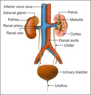
Anatomy of the Male Urinary Tract | Saint Luke's Health System Anatomy of the Male Urinary Tract. Your urinary tract helps to get rid of your body's liquid waste. The kidneys constantly filter the blood to collect unneeded chemicals and water, making urine. Urine travels through the ureters to the bladder. The bladder fills with urine, holding it until you're ready to release it. Signals from the brain ... Anatomy of the Urinary System | Johns Hopkins Medicine The organs of the urinary system include the kidneys, renal pelvis, ureters, bladder and urethra. The body takes nutrients from food and converts them to energy. After the body has taken the food components that it needs, waste products are left behind in the bowel and in the blood. Urinary System Label Worksheets & Teaching Resources | TpT Excretory System: Urinary Diagram to Label by Lori Maldonado 6 $2.00 PDF Students will add the provided labels to the diagram of the urinary system and then write a one sentence description of the function for each item. Labels include the bladder, ureter, urethra, kidney, dorsal aorta, vena cava, renal artery, and renal vein. PDF THE URINARY SYSTEM - University of Cincinnati The urinary system is composed of a pair of kidneys, a pair of ureters, a bladder, and a urethra. These components together carry out the urinary system's function of regulating the volume and composition of body fluids, removing waste products from the blood, and expelling the waste and excess water from the body in the form of urine.
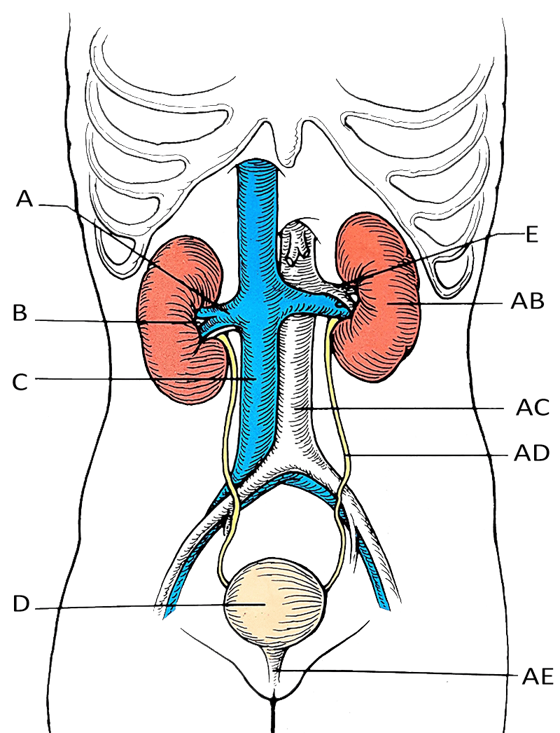
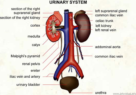
Urinary system quizzes and labeled diagrams | Kenhub Labeled diagram The best way to kick off your revision is with a urinary system diagram which clearly shows all of the structures found within. This gives you the opportunity to get a general feel of the appearance of each structure and their relations to the structures around them. Take a look at the urinary system diagram labeled below.
Urinary System Labeled Pictures, Images and Stock Photos Browse 44 urinary system labeled stock photos and images available, or start a new search to explore more stock photos and images. Newest results Labeled 3D Diagram of Female Reproductive System in Sagittal... Kidney Anatomy Labeled, Cross Section View on White Anatomy of cat with inside structure and organs scheme vector... The urinary system
Urinary System Diagram - Medical Art Library The urinary system is composed of the kidneys, ureters, bladder and urethra. Blood from the heart travels down the aorta where it enters the kidney via the renal arteries. The kidney acts as a filter and regulator, removing waste products (urea) and balancing glucose, electrolytes (salt, potassium and other minerals) and water levels in the ...
Urinary system: Organs, anatomy and clinical notes | Kenhub The urinary system consists of 4 major organs; the kidneys, ureters, urinary bladder and the urethra.Together these organs act to filter blood, remove waste products, create urine and transport urine out from the body. The urinary system is also called the excretory system, because held within the urine are the various excreted products, including by-products such as urea and uric acid, drugs ...

draw a well labelled diagram of the human urinary system - Biology - TopperLearning.com | 27g5awnxx
Urinary system Images, Stock Photos & Vectors | Shutterstock Find Urinary system stock images in HD and millions of other royalty-free stock photos, illustrations and vectors in the Shutterstock collection. Thousands of new, high-quality pictures added every day.
Urinary system diagram | How to draw labelled diagram of urin ... - YouTube How to draw Urinary system diagram. It is a labelled diagram of urin. Specially for class 9,10,11,12. QUE = WHAT IS URINARY SYSTEM ? ANS = .The Urinary Syste...
Labeled Urinary System Pictures, Images and Stock Photos CG image labeling the kidneys, adrenal glands, ureter, bladder and prostate gland inside a man's abdomen with other internal organs faded out on a white background. The urinary system The human urinary system medical illustration with internal organs Amoxicillin medication as international nonproprietary or... Flat vector illustration.
PDF The Urinary System - Pearson The organ system that performs this function in humans—the urinary system—is the topic of this chapter. The organs of the urinary system are organs of excretion—they remove wastes and water from the body. Specifically, the urinary system "cleans the blood" of metabolic wastes, which are substances produced by the body that it cannot
Urinary System Diagram - Kidney, Urinary Tract, Renal System Diagrams ... What is a Urinary System Diagram? Urinary system diagrams are illustrations of the urinary system, also referred to as the renal system. The urinary system, at a high level, contains two kidneys, two ureters, a urethra, and a bladder. The urinary system is located directly below the rib cage.
Urinary System Organs | Diagram, Structure & Anatomy - Video & Lesson ... The urinary system's major organs are the kidneys, ureter, urinary bladder, and urethra. This is also the urinary system order through which the urine flows out of the body. Gross Anatomy of the ...
Label and Color the Urinary System - The Biology Corner This simple worksheet asks students to label the major structures of the urinary system. They can also choose to color the diagram. I use coloring sheets in anatomy and physiology classes but this could also be used in biology or as a supplemental graphic for a frog or fetal pig dissection.
PDF HISTOLOGY OF URINARY SYSTEM - ndvsu.org Bowman'scapsule comprises of visceral and parietal layer. In between the two layers, Urinary space (US) is present which continues with the lumen of PCT. Visceral layer is lined by Podocytes.They have primary, secondary and tertiary processes, the smallest of these are called foot processes or pedicels. Narrow space between foot processes are called Filtration slits
Urinary System, Female, Anatomy - NCI Visuals Online The uterus is also shown. Anatomy of the female urinary system showing the kidneys, ureters, bladder, and urethra. Urine is made in the renal tubules and collects in the renal pelvis of each kidney. The urine flows from the kidneys through the ureters to the bladder. The urine is stored in the bladder until it leaves the body through the urethra.
17 Urinary System Labeling Diagram | Quizlet 17 Urinary System Labeling STUDY Learn Flashcards Write Spell Test PLAY Match Gravity Created by muskopf1TEACHER Terms in this set (7) Renal vein blood vessel that carries blood away from the kidney Kidney Filters waste from the blood like urea, water, salt and proteins. Ureter tube that carries urine from the kidney to the urinary bladder Aorta


Post a Comment for "39 diagram of urinary system with labels"