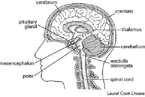
42 diagram of the brain with labels and functions
Ventricles of the Brain: Labeled Anatomy, Function, CSF Flow ... Learn the ventricles of the brain along with their definition, function, location, anatomy, and cerebrospinal fluid (CSF) flow using labeled diagrams. The ventricular system contains the lateral, third, and fourth ventricles whose function is to produce cerebrospinal fluid. Learn where CSF is found, The Human Brain: Anatomy and Function - Visible Body The brain directs our body's internal functions. It also integrates sensory impulses and information to form perceptions, thoughts, and memories. The brain gives us self-awareness and the ability to speak and move in the world. Its four major regions make this possible: The cerebrum, with its cerebral cortex, gives us conscious control of our ...
Major Structures and Functions of the Brain - NCBI Bookshelf Visual functions occupy the occipital lobe, the bulge at the back end of the brain. The primary area for visual perception is almost surrounded by the much larger visual association area. Nearby, extending into the lower part of the temporal lobe, is the association area for visual memory —a specialized area in the cortex.

Diagram of the brain with labels and functions
PDF Psychology Brain Structure/Anatomy and Function Psychology - Brain Structure/Anatomy and Function BRAIN FACTS Composition of the brain: 78% water, 12% lipids, 8% protein, 1% carbs, 2% soluble organics, and 1% salt ... Some products are also labeled incorrectly." Nicotine in e-cigarettes raise blood pressure. Compared to nonusers, users of e-cigarettes have a 71% higher risk of stroke, 59 ... Brain Anatomy Labeled Stock Illustrations - Dreamstime How the eye works medical scheme poster, elegant and minimal vector illustration, eye - brain labeled structure diagram. Stylized and artistic medical design. Inner organ icons vector illustration collection set. Labeled medical and anatomical human brain, lungs, heart, liver and stomach. ... Labeled diagram with location and functions. Frontal ... Brain Anatomy and How the Brain Works - Hopkins Medicine The cerebellum ("little brain") is a fist-sized portion of the brain located at the back of the head, below the temporal and occipital lobes and above the brainstem. Like the cerebral cortex, it has two hemispheres. The outer portion contains neurons, and the inner area communicates with the cerebral cortex.
Diagram of the brain with labels and functions. Structure of the Brain and Their Functions | New Health Advisor The function of these lobes is listed below: Frontal lobe- Associated with planning of speech, reasoning, emotions, problem solving and movement. Occipital lobe- It's associated with visual processing Parietal lobe- It's associated with recognition, movement, orientation, perception of stimuli, speech and memory. Human Brain Diagrams and Detailed Information - Innerbody Three layers of tissue, collectively known as the meninges, surround and protect the brain and spinal cord. The dura mater forms the leathery, outermost layer of the meninges. Dense irregular connective tissue made of tough collagen fibers gives the dura mater its strength. DOC Brain Anatomy Function Cheat Sheet Memory (remembering and learning) Amygdala Emotion (aggression) rage, fear Kluecer& Bucy Lesion monkey brain Hypothalamus Regulates thirst, hunger, body temperature, sexual behavior (hormone release). Controls/regulates maintenance reflexes (eating), Homeostasis linked to emotion. Helps govern endocrines. Monitors glands. Controls hunger. Labeled Brain Model Diagram - Science Trends The cerebrum is the largest and most complex portion of the human brain. The cerebrum's function is to control our actions and thoughts, either conscious or unconscious, and responses to stimuli. The cerebrum itself is typically divided into four different lobes: the temporal lobe, the parietal lobe, the occipital lobe, and the frontal lobe.
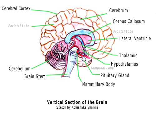
Lobes of the brain: Structure and function | Kenhub The cerebral cortex is a highly convoluted gray matter structure consisting of many gyri and sulci. The lobes of the cerebrum are actually divisions of the cerebral cortex based on the locations of the major gyri and sulci. The cerebral cortex is divided into six lobes: the frontal, temporal, parietal, occipital , insular and limbic lobes. Diagram of the Brain and its Functions - Bodytomy Given below is a diagram outlining the main brain functions and parts. Vertical Section of the Brain and its Functions Midbrain The midbrain is divided into two parts by the Aqueduct of Sylvius, which is the duct that connects the IIIrd ventricle in the midbrain with the IV ventricle in the pons and medulla oblongata. Diagram Of Brain with their Labelings and Detailed Explanation A well-labelled diagram of a human brain is given below for further reference. Structure And Function Of The Human Brain Parts Of The Human Brain The human brain is divided into three main parts: Forebrain. Midbrain. Hindbrain. These three main parts comprises many small parts. Forebrain The forebrain is also called as Prosencephalon. Parts of the brain: Learn with diagrams and quizzes | Kenhub Labeled brain diagram First up, have a look at the labeled brain structures on the image below. Try to memorize the name and location of each structure, then proceed to test yourself with the blank brain diagram provided below. Labeled diagram showing the main parts of the brain Blank brain diagram (free download!)
Label the Brain Anatomy Diagram Flashcards | Quizlet Start studying Label the Brain Anatomy Diagram. Learn vocabulary, terms, and more with flashcards, games, and other study tools. Home. Subjects. Explanations. Create. ... Brain Anatomy and Function. 52 terms. kmgeary2395. Labeling Brain and Ventricles. 19 terms. mkcoleman PLUS. Other sets by this creator. Social Development Final. 84 terms. Parts of the Brain Activity for Kids, Brain Diagram, and Worksheets for ... Their are 2 brain function worksheets where your student will learn about the different parts of the brain your child will learn about are: FRONTAL LOBES - The frontal lobes control voluntary movement such as reasoning, planning, parts of speech and movement, emotions, and problem-solving It is fully developed by age 10. DOC Label the Brain Anatomy Diagram Answers: Label the Brain Diagram The Brain. Read the definitions below, then label the brain anatomy diagram. Cerebellum - the part of the brain below the back of the cerebrum. It regulates balance, posture, movement, and muscle coordination. Corpus Callosum - a large bundle of nerve fibers that connect the left and right cerebral hemispheres. The Brain - Diagram and Explanation A diagram of how the brain works From Building Mental Muscle Glossary of Terms Six Brain Functions AMYGDALA: Lying deep in the center of the limbic emotional brain, this powerful structure, the size and shape of an almond, is constantly alert to the needs of basic survival including sex, emotional reactions such as anger and fear.
brain diagrams
Anatomy of the Brain - Simply Psychology The temporal lobes are located on both sides of the brain, near the temples of the head, hence the name temporal lobes (Figure 5). The main functions of these lobes include understanding, language, memory acquisition, face recognition, object recognition, perception, and processing auditory information.
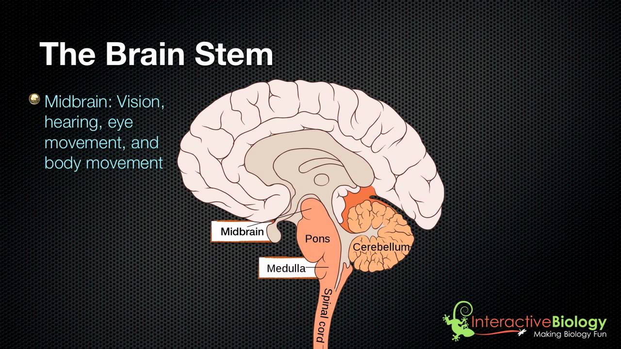
027 The 3 parts of the brain stem and their functions - YouTube
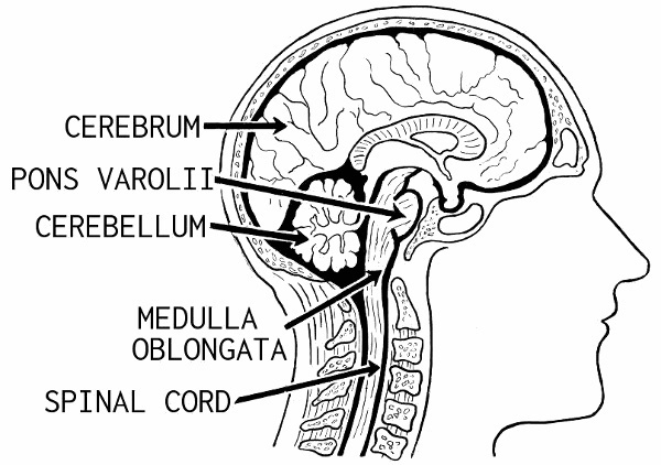
Human Brain: Structure, Location, Function, Parts & Pictures Spinal cord is the central nervous system component that begins in the lower area of the brain, extending along the spine. The spinal cord links the brain with the nerves. The spinal cord's nerve tissues are approximately 45 centimeters long, and nearly 2 centimeters bulk, and they conform the peripheral nervous system. Function of the Brain

Modularity in the Nervous System
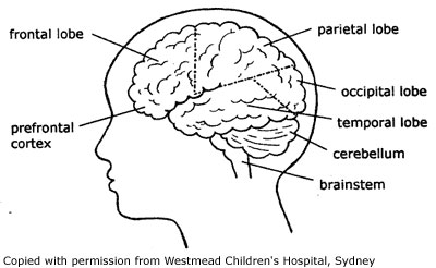
Brain (Human Anatomy): Picture, Function, Parts, Conditions, and More • The cerebellum is at the base and the back of the brain. The cerebellum is responsible for coordination and balance. The brain is also divided into several lobes: • The frontal lobes are...
Brain Diagram - Cliparts.co
Nervous System - Label the Brain - TheInspiredInstructor.com This brain part controls balance, movement, and coordination. (11) This brain part controls involuntary actions such as breathing, heartbeats, and digestion. (12) This part of the nervous system moves messages between the brain and the body. (13) This part of the cerebrum interprets and sorts information from the senses. (14)

12 best images about BRAIN FUNCTIONS on Pinterest | Brain anatomy, Medical illustration and Medical
Label The Brain Psychology Teaching Resources | Teachers Pay Teachers This bundle pack includes a 2-hour comprehensive presentation on the parts and functions of the brain. It also includes a student-guided notes document where they label a diagram of the brain. It also includes a master copy of that diagram for grading. Subjects: Psychology, Social Studies - History. Grades:

Label The Brain Anatomy Diagram Answers - Pensandpieces
Structure, Diagram, Parts Of Human Brain - BYJUS The hypothalamus is a small and essential part of the brain, located precisely below the thalamus. It is considered the primary region of the brain, as it is involved in the following functions: Receives impulses Regulates body temperature Controls the mood and emotions Controls the sense of taste and smell Synthesises the body's essential hormones

Brain Function
Anatomy of the Brain: Structures and Their Function The forebrain is the division of the brain that is responsible for a variety of functions including receiving and processing sensory information, thinking, perceiving, producing and understanding language, and controlling motor function. There are two major divisions of forebrain: the diencephalon and the telencephalon.
Kids Health Information : Brain injury - How the brain works
Parts of the Human Brain | Anatomy & Function - Study.com The parts of the brain include the cerebrum, the cerebellum, the brain stem, and the pituitary gland. The brain structure is protected by the skull, which is composed of the cranium and the bones...

cerebral cortex | Brain diagram, Human brain diagram, Brain anatomy
Labeled Diagrams of the Human Brain You'll Want to Copy Now Labeled Diagrams of the Human Brain Central Core The central core consists of the thalamus, pons, cerebellum, reticular formation and medulla. These five regions are the central areas that regulate breathing, pulse, arousal, balance, sleep and early stages of processing sensory information.
brain diagram 2 - /medical/anatomy/brain/brain_diagram_2.png.html
Lobes of the brain - Queensland Brain Institute The brain's cerebral cortex is the outermost layer that gives the brain its characteristic wrinkly appearance. The cerebral cortex is divided lengthways into two cerebral hemispheres connected by the corpus callosum. Traditionally, each of the hemispheres has been divided into four lobes: frontal, parietal, temporal and occipital . (Wikimedia)
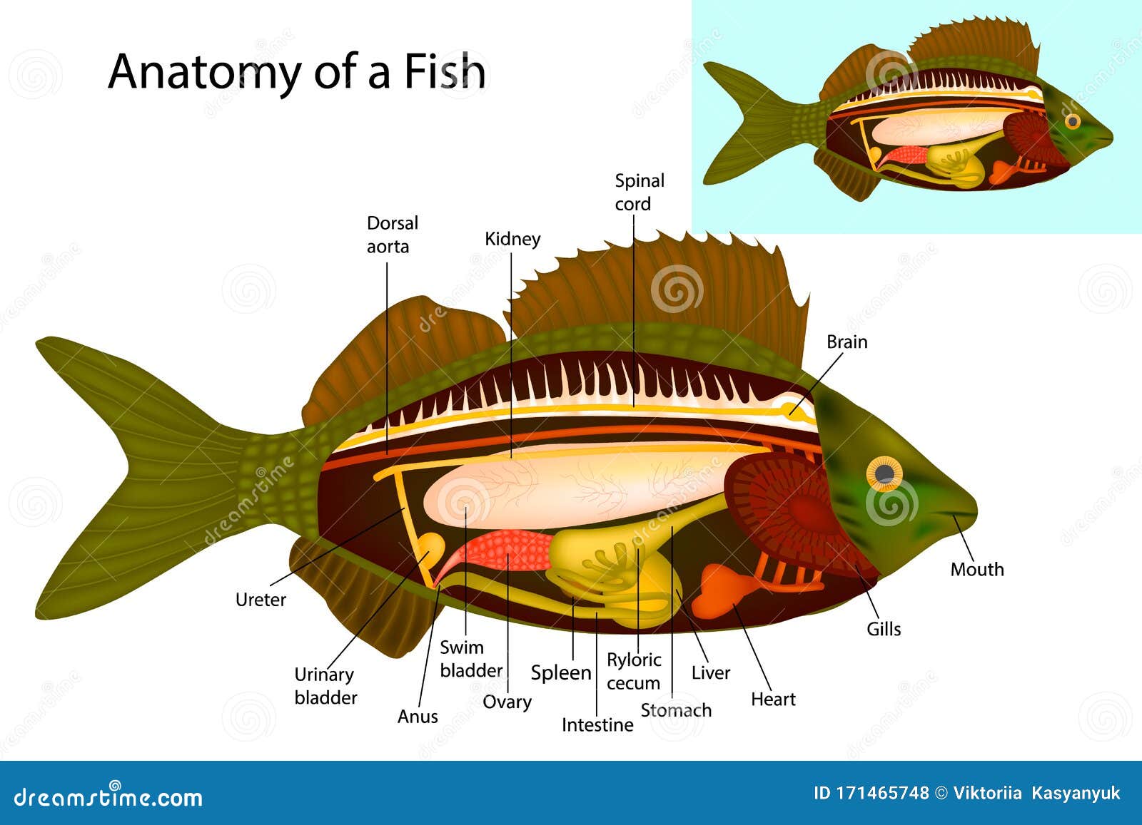
Fish Internal Organs Vector Art Diagram Anatomy Without Labels | CartoonDealer.com #72048931
Brain Anatomy and How the Brain Works - Hopkins Medicine The cerebellum ("little brain") is a fist-sized portion of the brain located at the back of the head, below the temporal and occipital lobes and above the brainstem. Like the cerebral cortex, it has two hemispheres. The outer portion contains neurons, and the inner area communicates with the cerebral cortex.
32 Blank Brain Diagram To Label - Labels Database 2020
Brain Anatomy Labeled Stock Illustrations - Dreamstime How the eye works medical scheme poster, elegant and minimal vector illustration, eye - brain labeled structure diagram. Stylized and artistic medical design. Inner organ icons vector illustration collection set. Labeled medical and anatomical human brain, lungs, heart, liver and stomach. ... Labeled diagram with location and functions. Frontal ...
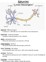
Little messengers - WikiEducator
PDF Psychology Brain Structure/Anatomy and Function Psychology - Brain Structure/Anatomy and Function BRAIN FACTS Composition of the brain: 78% water, 12% lipids, 8% protein, 1% carbs, 2% soluble organics, and 1% salt ... Some products are also labeled incorrectly." Nicotine in e-cigarettes raise blood pressure. Compared to nonusers, users of e-cigarettes have a 71% higher risk of stroke, 59 ...

Found on Bing from www.pinterest.com | Brain anatomy and function, Brain diagram, Brain anatomy

Educative diagrams: The Female Reproductive System
Post a Comment for "42 diagram of the brain with labels and functions"