40 picture of the eye with labels
Eye Anatomy: 16 Parts of the Eye & Their Functions The lens of the eye (or crystalline lens) is the transparent lentil-shaped structure inside your eye. This is the natural lens. It is located behind the iris and to the front of the vitreous humor (vitreous body). The vitreous humor is a clear, colorless, gelatinous mass that fills the gap between the lens and the retina in the eye. Human eye anatomy Images, Stock Photos ... - Shutterstock 60,203 human eye anatomy stock photos, vectors, and illustrations are available royalty-free. See human eye anatomy stock video clips Image type Orientation Sort by Popular Biology Healthcare and Medical Icons and Graphics Recreation/Fitness human eye anatomy 3d rendering eye visual perception infographic Next of 603
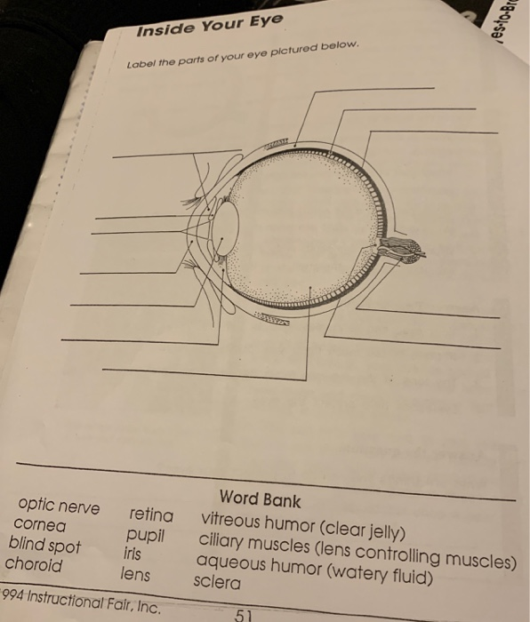
Human Eye Coloring Page | crayola.com Human Eye. Use Crayola® crayons, colored pencils, or markers to color the parts of the human eye. Use the word bank below to identify parts of the eye.The eye is the organ that collects images and sends them to the brain, so you can see. The eye is protected by the bones of your skull and six muscles.
Picture of the eye with labels
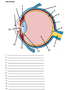
Label the Eye - The Biology Corner Label the Eye. Shannan Muskopf December 30, 2019. This worksheet shows an image of the eye with structures numbered. Students practice labeling the eye or teachers can print this to use as an assessment. There are two versions on the google doc and pdf file, one where the word bank is included and another with no word bank for differentiation. FREE! - Label the Eye Worksheet - Teacher-Made Learning ... In this resource, you'll find a 2-page PDF that is easy to download, print out, and use immediately with your class. The first page is a labelling exercise with two diagrams of the human eye. One is a view from the outside, and the other is a more detailed cross-section. On the second page, you'll find a set of answers showing the properly labelled human eyes, designed to help you check ... Human Eye Diagram - Human Body Pictures & Images - Science ... Photo description: This human eye diagram gives an excellent overview of the human eye. The cross section features labeled parts such as the iris, pupil, cornea, lens, retina, choroid, optic disc, optic nerve and fovea. For more information on eyes, check out our range of interesting human eye facts.
Picture of the eye with labels. Eye Diagram Unlabelled - Wiring Diagram Pictures 07.12.2018. 1 Comments. on Eye Diagram Unlabelled. Select the correct label for each part of the eye. The image is taken from above the left eye. Click on the Score button to see how you did. Incorrect answers will. 61 high-quality Unlabeled Eye Diagram for free! human eye diagram. YouTube. Eye anatomy: A closer look at the parts of the eye Eye anatomy: A closer look at the parts of the eye. By Liz Segre. When surveyed about the five senses — sight, hearing, taste, smell and touch — people consistently report that their eyesight is the mode of perception they value (and fear losing) most. Despite this, many people don't have a good understanding of the anatomy of the eye, how ... Transverse Section Of Eye Anatomy With Labels High-Res ... Transverse section of eye anatomy with labels. - stock illustration. Transverse section of eye anatomy with labels. Buy the print. Get this image in a variety of framing options at Photos.com. Printable Eye Images | Etsy Vintage image Human Eye Instant Download picture Digital printable clipart graphic Burlap Fabric Transfer Iron On Decor T-shirt 300dpi. Ad by UnoPrint Ad from shop UnoPrint. UnoPrint. From shop UnoPrint. 5 out of 5 stars. (1,869) Sale Price $1.96. $1.96. $2.80.
Eye Anatomy Diagram - EnchantedLearning.com Definitions : Aqueous humor - the clear, watery fluid inside the eye. It provides nutrients to the eye. Astigmatism - a condition in which the lens is warped, causing images not to focus properly on the retina. Binocular vision - the coordinated use of two eyes which gives the ability to see the world in three dimensions - 3D. CUT-AND-ASSEMBLE PAPER EYE MODEL • thin permanent marker for a number labels on plastic parts (such as a very thin point Sharpie) Assembly: 1) After copying pattern pages onto card stock, cut out all parts. On the background page that says THE HUMAN EYE, cut away the black rectangles and trim the triangles at the bottom, as shown in picture above. The Eye - Science Quiz - Seterra The Eye - Science Quiz: Our eyes are highly specialized organs that take in the light reflected off our surroundings and transform it into electrical impulses to send to the brain. The anatomy of the eye is fascinating, and this quiz game will help you memorize the 12 parts of the eye with ease. Light enters our eyes through the pupil, then passes through a lens and the fluid-filled vitreous ... PDF Eye Anatomy Handout - National Eye Institute of light entering the eye. Lens: The lens is a clear part of the eye behind the iris that helps to focus light, or an image, on the retina. Macula: The macula is the small, sensitive area of the retina that gives central vision. It is located in the center of the retina. Optic nerve: The optic nerve is the largest sensory nerve of the eye.
20 Different Ways to Draw the Eye - Improve Drawing Realistic Eye Drawings. Creating a realistic drawing of an eye is a technique that will enable you to break down the eye's form to its basic parts.. This way of drawing the eye is best begun with a faintly drawn line. Draw the eye from direct observation using a mirror or drawing a model. Eye Diagram - Differentiated Worksheets and EASEL Activities Jan 29, 2016 - Use these simple eye diagrams to help students learn about the human eye. Three differentiated worksheets are included: 1. Write the words using a word bank2. Cut and paste the words3. Write the words without a word bank Labels include: eyebrow, eyelid, eyelashes, pupil, iris, and sclera.UPDATE:I'... Human Eye Anatomy Pictures, Images and Stock Photos Browse 7,999 human eye anatomy stock photos and images available, or search for vision or retina to find more great stock photos and pictures. Newest results vision retina human eye structure eye chart human eyeball eye doctor eye diagram cataract retinopathy pancreas Anatomy of human eye and descriptions. Labelling the eye - Science Learning Hub In this interactive, you can label parts of the human eye. Use your mouse or finger to hover over a box to highlight the part to be named. Drag and drop the text labels onto the boxes next to the eye diagram If you want to redo an answer, click on the box and the answer will go back to the top so you can move it to another box.
33 Label The Parts Of The Eye - Labels For You
PDF Parts of the Eye - National Eye Institute Parts of the Eye . To understand eye problems, it helps to know the different parts that make up the eye and the functions of these parts. Here are descriptions of some of the main parts of the eye: Cornea: The cornea is the clear outer part of the eye's focusing system

Label Eye Diagram
Label Parts of the Human Eye - University of Dayton Parts of the Eye Select the correct label for each part of the eye. The image is taken from above the left eye. Click on the Score button to see how you did. Incorrect answers will be marked in red.

World's Beautiful things around us !: Beautiful nature| Eye cooling pictures every body loves to ...
blank eye diagrams - Bing Images | Human eye diagram ... Label the muscles of the arm. Designed for an anatomy class, two images of the arms can be colored and labeled, one focusing on the flexors and the other on the extensors. Georgina Miller

Melanie Hicks Hot Pics and Bio | Picture Perfect
What is an eye mark and why do I need it? - Consolidated Label An 'eye mark' (also known as 'eye spot') is a small rectangular printed area located near the edge of the printed flexible packaging material. A sensor on the form-fill-seal (FFS) machine reads the eye mark to identify packaging material, control the material's position, and coordinate the separation and cutting of the flexible packaging material.

Missing Beats of Life: Eyes HD Wallpapers and Images
The Eye - diagram to label | Teaching Resources File previews. pdf, 2.94 MB. Diagram of eye with key words to use in labelling it. Tes classic free licence.
30 Label Eye - Labels For Your Ideas
The Human Eye (Eyeball) Diagram, Parts and Pictures The eyeball is a round gelatinous organ that contains the actual optical apparatus. It is approximately 25 mm in diameter and sits snugly in the orbit where six muscles control its movement. The eyeball has three layers, each of which has several important structures that are essential for the sense of vision. Wall of the Eyeball

Street Art By ArtFlyMovie: NYCHOS THE WEIRD - Some Extracts of his Amazing Work
Structure and Functions of Human Eye with labelled Diagram Human Eye Diagram: Contrary to popular belief, the eyes are not perfectly spherical; instead, it is made up of two separate segments fused together. Explore: Facts About The Eye To understand more in detail about our eye and how our eye functions, we need to look into the structure of the human eye.
Eye With Labels clip art (111109) Free SVG Download / 4 Vector
500+ Stickers for Kids | Oriental Trading Company Stickers aren't just for kids! Use fun stickers for scrapbooking and create a special picture album, personalize them for party favors or wedding favors, place labels to special items or add them to your craft supplies. You can even add a sticker to your greeting cards or add a cute one as an envelope seal. Whatever you decide to create or use ...

Label an Eye
Label Eye Printout - EnchantedLearning.com Label the Eye Diagram. Human Anatomy. Read the definitions, then label the eye anatomy diagram below. Cornea - the clear, dome-shaped tissue covering the front of the eye. Iris - the colored part of the eye - it controls the amount of light that enters the eye by changing the size of the pupil. Lens - a crystalline structure located just behind ...
+(Left+Eye)+(2).jpg)
Aquarious' TLC Fan Blog: TLC's 'Waterfalls' Video Photo Shots
Eye Diagram With Labels and detailed description - BYJUS A brief description of the eye along with a well-labelled diagram is given below for reference. Well-Labelled Diagram of Eye The anterior chamber of the eye is the space between the cornea and the iris and is filled with a lubricating fluid, aqueous humour. The vascular layer of the eye, known as the choroid contains the connective tissue.
Aquarious' TLC Fan Blog: TLC's 'Waterfalls' Video Photo Shots
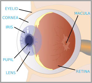
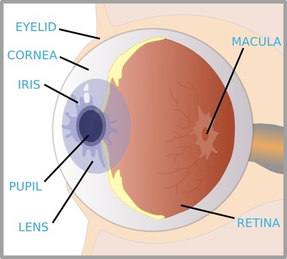
Eye Anatomy Detail Picture Image on MedicineNet.com Picture of Eye Anatomy Detail The eye is our organ of sight. The eye has a number of components which include but are not limited to the cornea, iris, pupil, lens, retina, macula, optic nerve, choroid and vitreous. Cornea: clear front window of the eye that transmits and focuses light into the eye.

Eclectitude: Bleigiessen Glass Sculpture - Thomas Heatherwick, Artist
A Picture of the Eye - WebMD One eye sees better than the other, so your brain favors that eye. The weaker eye, which may or may not wander, is called the "lazy eye." Astigmatism : A problem with the curve of your cornea.

Eye Label
Solved B с A E F D Match the following parts of the eye ... Science. Anatomy and Physiology. Anatomy and Physiology questions and answers. B с A E F D Match the following parts of the eye with the labels in the picture above. A Iris F Cornea В. Ciliary Muscles G Optic Nerve C Lens E Retina Aqueous and Vitreous Fluid. Question: B с A E F D Match the following parts of the eye with the labels in the ...

The BioLogs: CSEC - The Eye - functions of the various parts
Human Eye Diagram - Human Body Pictures & Images - Science ... Photo description: This human eye diagram gives an excellent overview of the human eye. The cross section features labeled parts such as the iris, pupil, cornea, lens, retina, choroid, optic disc, optic nerve and fovea. For more information on eyes, check out our range of interesting human eye facts.
Vector label for free download about (6,449) vector label. sort by newest first page (95/95)
FREE! - Label the Eye Worksheet - Teacher-Made Learning ... In this resource, you'll find a 2-page PDF that is easy to download, print out, and use immediately with your class. The first page is a labelling exercise with two diagrams of the human eye. One is a view from the outside, and the other is a more detailed cross-section. On the second page, you'll find a set of answers showing the properly labelled human eyes, designed to help you check ...

Sports Day / Fun Run Poster | Free Early Years & Primary Teaching Resources (EYFS & KS1)
Label the Eye - The Biology Corner Label the Eye. Shannan Muskopf December 30, 2019. This worksheet shows an image of the eye with structures numbered. Students practice labeling the eye or teachers can print this to use as an assessment. There are two versions on the google doc and pdf file, one where the word bank is included and another with no word bank for differentiation.
Post a Comment for "40 picture of the eye with labels"